Leg Muscles Diagram Front / Labeled Muscles Of Lower Leg Yahoo Search Results Leg Muscles Anatomy Lower Leg Muscles Front Leg Muscle - The radial nerve in particular is a nerve that can be predisposed to several problems such as dogs having trouble moving their front legs and possibly developing muscle wasting.
Leg Muscles Diagram Front / Labeled Muscles Of Lower Leg Yahoo Search Results Leg Muscles Anatomy Lower Leg Muscles Front Leg Muscle - The radial nerve in particular is a nerve that can be predisposed to several problems such as dogs having trouble moving their front legs and possibly developing muscle wasting.. A muscle along the outside of the leg that bends the foot out at the ankle. The hip muscles work together to carry out 4 different types of movement: The muscles in the hip are responsible for the movement of the hip and, by proxy, the leg. Together, these muscles straighten your knee, stabilize your knee joint, assist in flexing your hip (drawing your knee towards your chest), and help absorb force when you land after jumping or leaping. The quad muscles— which form the meaty mass on the front of your thighs — are among your strongest muscle groups, and play a critical role in athletic activities.
It lies between the knee and the ankle, while the upper leg. Metatarsal # 1 (big toe) raises front of. This chart is beautifully illustrated and offers the most comprehensive look at the muscles of the human leg available. The following diagram illustrates the actions of the terms adduction, abduction, flexion and extension at the different joints. 6 / 10 ( 2 votes ) muscle of the human leg diagram.
Muscle Diagram Skeletal Muscles from www.changingshape.com A muscle located on the back portion of the lower leg, being one of the two major muscles that make up the calf:the flexing of this muscle during walking and bending of the knee creates traction on the femur, pulling it toward the tibia in the lower leg and causing the knee to bend. This important tendon in the back of the calf and ankle stores the elastic energy needed for running, jumping, and other physical activity. The radial nerve in particular is a nerve that can be predisposed to several problems such as dogs having trouble moving their front legs and possibly developing muscle wasting. Together with the upper leg, it forms the lower extremity. Human anatomy for muscle, reproductive, and skeleton. The hip muscles work together to carry out 4 different types of movement: They work to straighten or extend your leg. The hamstrings are three muscles at the back of the thigh that affect hip and knee movement.
The muscle groups can work independently for specific movements.
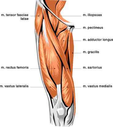
It is also visible on the medial edge of the thigh from the anterior. In this image, you will find muscle of the human leg diagram, hip and femur middle layer, hip and femur deep layer, overview of the most important muscles of the leg, femur middle layer, femur deep layer, rectus femoris m. Front view of human body: The muscles in the back help with. The lower leg is a major anatomical part of the skeletal system. These four muscles at the front of the thigh are the major extensors of the knee. Anatomical diagram of internal organs. 6 / 10 ( 2 votes ) muscle of the human leg diagram. Anterior muscles of the lower leg and their functions. Calcaneum (by achilles tendon) raises heal when leg is bent. Soleus (calf muscles) tibia and fibula: These four muscles at the front of the thigh are the major extensors (help to extend the leg. This involves pointing the toes upward.
Anatomical diagram of internal organs. 10 / 10 ( 1 vote ) anatomy of the groin area superficial muscles and deep muscles. This high level of complexity is. It gets its blood flow from the arteries in the tiberial artery. The four muscles that make up the quadriceps are the strongest and leanest of all muscles in the body.these muscles at the front of the thigh are the major extensors (help to extend the leg.
Pin On Health Metabolism from i.pinimg.com Posterior compartment, also known as the flexor compartment; This is why you have to indicate which biceps you are taking about when discussing one or other of these muscles. This muscle diagram is interactive: Together with the upper leg, it forms the lower extremity. A muscle along the outside of the leg that bends the foot out at the ankle. This involves pointing the toes upward. These four muscles at the front of the thigh are the in this image, you will find muscle of the human leg diagram, hip and femur middle layer, hip and femur deep layer, overview of the most important. The muscles involved include the rectus femoris, vastus intermedius, vastus medialis (inner thigh), and vastus lateralis (outer thigh).
Pulls back scapula (shoulder blades).
Quadriceps muscle anatomy 3d medical illustration. Muscles of the anterior hip and thigh. The muscles in the front allow for dorsiflexion. In this image, you will find muscle of the human leg diagram, hip and femur middle layer, hip and femur deep layer, overview of the most important muscles of the leg, femur middle layer, femur deep layer, rectus femoris m. This is the biggest muscle that is in the tibialis anterior. A muscle located on the back portion of the lower leg, being one of the two major muscles that make up the calf:the flexing of this muscle during walking and bending of the knee creates traction on the femur, pulling it toward the tibia in the lower leg and causing the knee to bend. On the medial edge of the posterior thigh is the gracilis muscle. #front leg muscle diagram #leg muscle diagram wikipedia #muscle anatomy of leg and foot #muscle diagram of upper leg #muscular diagram of leg. Movements like and take some extend one leg straight out in front of you, balancing on your other foot. Legs are used for standing, and all forms of. These four muscles at the front of the thigh are the major extensors of the knee. Some common causes of leg pain include: The basic function of the quadriceps muscle is to extend and straighten the leg.
For a more detailed anatomy of the muscle, check out the following leg muscle diagrams posted below. The muscles involved include the rectus femoris, vastus intermedius, vastus medialis (inner thigh), and vastus lateralis (outer thigh). Notice the upper leg has a biceps muscle just like the upper arm does. Muscle system diagram 12 photos of the muscle system diagram muscular system diagram blank, muscular system diagram front and back, muscular system diagram printable, muscular system key diagram 2, printable muscular system diagram download, human muscles, muscular system diagram blank, muscular system diagram front and. This muscle is one of the ones that help.
Human Anatomy Fundamentals Muscles And Other Body Mass from cdn.tutsplus.com The human leg, in the general word sense, is the entire lower limb of the human body, including the foot, thigh and even the hip or gluteal region. These muscles pull the toes and feet upward, a process known as dorsiflexion. Quadriceps muscle anatomy 3d medical illustration. It is located on the lateral side of the tibia and is the largest muscle in the anterior portion of the leg. The quadriceps muscles are the large muscles that make up the front of the thighs. You may also find vastus intermedius deep layer, vastus medial. Front leg injuries are usually easy to deal with as the dog tends to put more weight on the rear legs especially while running. They also help in dorsiflexion.
The fibularis longus originates from the head and upper lateral surface of the fibula, runs in a bony groove along the bottom of the foot to attach on the other side at the base of the first metatarsal and the neighboring medial cunieform bone, and acts to evert the.
Anatomical diagram of internal organs. The tibialis anterior also assists in turning the foot inward. The fibularis longus originates from the head and upper lateral surface of the fibula, runs in a bony groove along the bottom of the foot to attach on the other side at the base of the first metatarsal and the neighboring medial cunieform bone, and acts to evert the. You may also find transversus abdominis, iliopsoas, gluteus medius, pectineus, adductor longus. 6 / 10 ( 2 votes ) muscle of the human leg diagram. Most leg pain results from wear and tear, overuse, or injuries in joints or bones or in muscles, ligaments, tendons or other soft tissues. In this image, you will find muscle of the human leg diagram, hip and femur middle layer, hip and femur deep layer, overview of the most important muscles of the leg, femur middle layer, femur deep layer, rectus femoris m. Anterior compartment, also known as the extensor compartment; It gets its blood flow from the arteries in the tiberial artery. It is also visible on the medial edge of the thigh from the anterior. This high level of complexity is. Metatarsal # 1 (big toe) raises front of. The muscles work together to enable movement and keep the hip in alignment.
You may also find vastus intermedius deep layer, vastus medial leg muscles diagram. The muscles in the back help with.


0 Komentar